Page History
caMicroscope is a tool to view, annotate, and analyze whole-slide, biomedical images. It manages digital pathology images, associated clinical and imaging metadata, and human/machine generated annotations and markups. It also allows you to view and analyze nuclear segmentations of images.
This guide explains how to do the following tasks in caMicroscope:
...
- Use the Chrome browser to navigate to caMicroscope.
The Sign In page appears.
- Use your Google credentials to sign in to caMicroscope.
The caMicroscope site appears.
- Select one of the image databases by clicking either the large slide image or the More button. Options include the Breast Cancer Genomic Pilot, Virtual Tissue Repository (VTR) Pending Slides, and Pancreatic Ductal Adenocarcinoma (PDAC).
A table appears that lists all whole-slide images for the selected database that you are authorized to view.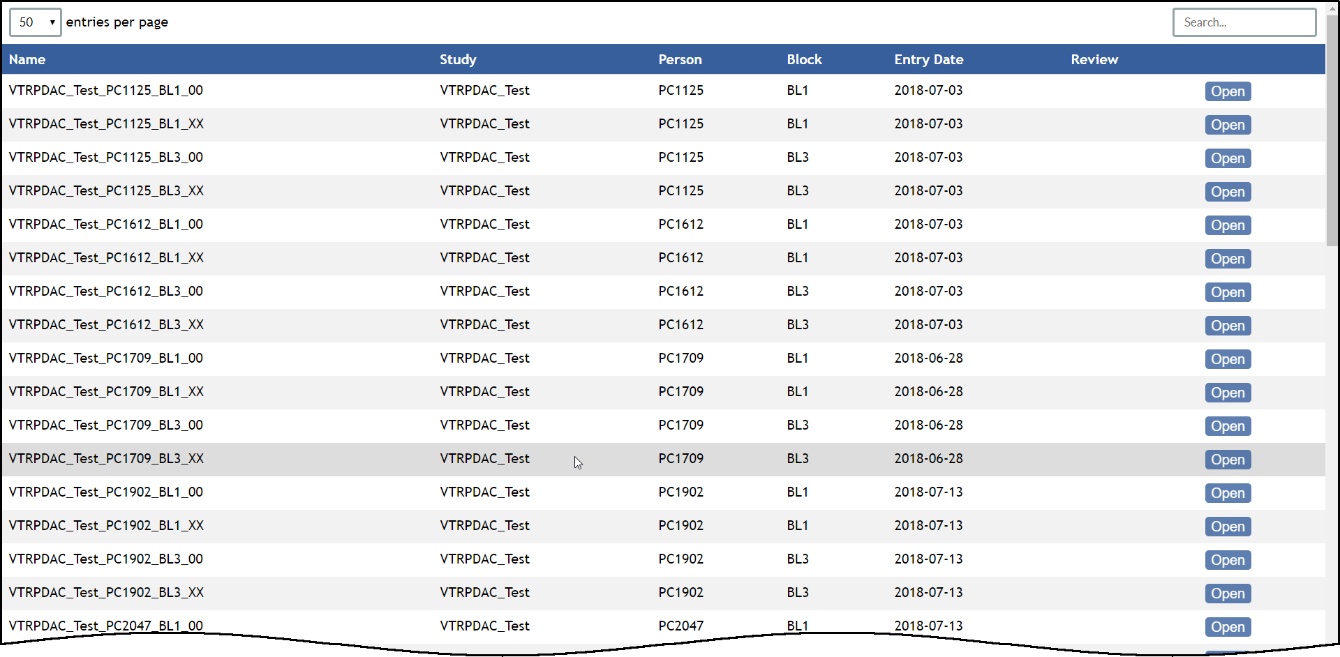
In the table, click the Open button for any row.
The slide opens in caMicroscope.Note To select a different image, click
 in the caMicroscope toolbar or the back arrow in your browser to return to the table.
...